
|
||||||||||||||||
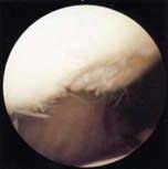 |
| FIGURE 1 - Arthroscopic photograph of fairly advanced chondromalacia afflicting the gliding surface cartilage behind the patella (top section of photo). Unlike what you see here, the surface of the patella is normally quite smooth. |
If patellar cartilage deterioration proceeds beyond simple softening and involves fissuring, disintegration and/or outright erosion (see FIGURE 1), the joint surface can become quite irregular and a sensation of grinding or catching may be felt by the patient when extending (straightening) the knee. This is often more pronounced when ascending stairs, or when getting back up from a bent-knee (squat) position. Such symptoms can often be improved by surgery.
During an arthroscopic "chondroplasty" procedure the roughened and degenerated articular cartilage tissue behind the patella is shaved down, re-contoured and evened out to the maximum extent possible or practical, using arthroscopic instruments (see FIGURE 2). Laser or radio frequency (r.f.) electro-thermal ablation "sculpting" techniques can also be employed, but extreme care must be taken not to allow excessive heat build-up within nearby healthy cartilage or the underlying bone, as this can cause localized cartilage coagulation, or worse, bone death (avascular necrosis), with potentially catastrophic results. I once encountered a patient treated by another surgeon, in whom a simple, outpatient laser chondroplasty resulted in enough bone death within the patella to ultimately require its complete surgical removal (patellectomy)!
 |
|
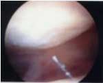
FIGURE 2 - Photograph of a deteriorated patellar surface after arthroscopic shaving. While not restored to normal, a firmer, more even gliding surface has been created and the patient should have less pain and experience less patellar grinding/catching sensations. |
If arthroscopy is performed to treat a deteriorated patellar
articular cartilage surface, concomitant surgical release (division)
of the lateral retinaculum is often (but not always) appropriate.
A retinacular release can be done from inside the knee
using an arthroscope. In cases of extensive chondromalacia combined
with poor patellar alignment and/or patellar instability, extensive
patellar realignment surgery is sometimes needed. This may involve
lower thigh muscle/tendon transfers and/or some "carpentry"
on the upper tibia ("shin bone") to redirect the angle
at which the patellar tendon pulls on the patella.
After patellar surgery a patient's knee and leg may temporarily behave as if they are "in shock". The knee joint may remain swollen and irritable for quite some time, and the leg may lose quite a bit of thigh muscle tone and strength. Sometimes special physical therapy regimens that involve electrical muscle stimulation and biofeedback are required to gradually "re-educate" the dysfunctional leg muscles, teaching them how to contract fully again so that they may then be strengthened through routine resistance exercise. Despite such difficulties, if a patient's knee pain is alleviated by the surgery, near-complete restoration of muscle function may be possible following a diligent rehabilitation regimen.
Arthroscopic treatment of chondromalacia patella, with or without
lateral retinacular release or other more major procedures, can
produce gratifying results, but does not represent a total "cure"
for the problem. Once articular cartilage has become degenerated,
simply shaving away the worst of it does not really restore a
joint to normalcy. Sometimes the procedure is palliative, at best,
and subsequent surgery and/or specialized brace treatment may
be needed. Despite recent advances in surgery, physical therapy,
medications and knee bracing, the problem of the stubbornly painful
patellofemoral joint is one that orthopedic surgeons have yet
to solve, short of prosthetic joint replacement.
|
|
||
|
While patellar pain problems are common, they can easily be mismanaged. Knee specialty care is always a good idea! If you have questions concerning your own or a family member's knee condition, you are invited to have those questions answered by way of a consultation here at The Knee and Shoulder Centers. We have anatomic charts, models, and educational videotapes available to help you participate actively in the medical assessment and decision-making process. |
||
|
|
||
|
|
||
|
|
|
|
|
|