"Chondromalacia Patella" in non-technical terms, means "soft kneecap cartilage". While everyone develops chondromalacia of their joints to one degree or another as they age, the articular (joint surface) cartilage that lines the back of the patella seems particularly prone to gradual deterioration and breakdown. The patella is often the first bone in the body to show aging (degenerative) change, beginning in most of us by the age of 35. Clinical evidence indicates that this natural degeneration can be accelerated by genetic factors, obesity, unusually stressful and repetitive use of the patellofemoral joint, and/or structural joint injury. In some individuals, chondromalacia patella can occur as early as the teenage years. Such premature chondromalacia is seen somewhat more frequently in females than in males. While treatment is available for this condition, there is no universally successful remedy. We never seem to pay attention to our kneecaps until they begin to hurt! Chondromalacia is probably the single most common condition suffered by our kneecaps, sometimes causing pain and sometimes not. "Creaky" kneecaps are generally afflicted with chondromalacia, but medical studies have shown that people with such noisy kneecaps rarely notice anything abnormal about them until they actually become painful. Congenital (inborn) problems with a person's knees such as patellar malalignment (a natural imbalance of patellar posture) can cause chondromalacia by early adulthood. Malalignment affects patellar movement patterns and causes uneven stress to be placed on the patellar joint surface, perhaps similar to the abnormal pressure and friction that produces the uneven wear seen on automobile tires that are out of alignment. Over time, this may lead to advanced patellofemoral arthritis, occasionally occurring as early as the fourth decade of life. Chronic patellar overloading caused by obesity and/or many years of repetitive knee bending/squatting also seems to be associated with premature patellar cartilage breakdown, even in knees with normal patellar alignment. Chondromalacia patella (which can be considered an early stage of localized,
knee joint arthritis) will sometimes remain clinically silent (cause
no symptoms) for many years, whereas in other cases it becomes annoying
and obvious early on. Patellar pain, combined with crepitation (a sensation
of grinding, catching or creaking as one moves the knee), is typical.
Sometimes inflammation causes fluid and swelling to develop in the knee.
Activities such as stair climbing, squatting and kneeling often become
uncomfortable, and in rare cases, almost impossible. DiagnosisChondromalacia patella should not be confused with simple patellar arthralgia ("achy kneecap syndrome", for lack of a better common name), which causes a dull, achy pain within the kneecap bone itself. This pain makes it difficult to keep one's knee flexed (bent) for prolonged periods, such as in movie theaters or airplanes. While this syndrome can co-exist with patellar chondromalacia, it can cause symptoms even when the articular cartilage behind the kneecap is perfectly firm and healthy. Patellar arthralgia, by itself, is usually recognizable as such by knee specialists. It is usually best treated with therapeutic exercise alone, emphasizing quadriceps muscle strengthening and stretching. Surgery rarely offers any benefit. When knee pain is caused by structural softening and degeneration of the articular cartilage behind the kneecap (true chondromalacia), the patient's orthopedic surgeon should search for underlying mechanical causes such as natural patellar malalignment, a history of recurrent injury caused by unstable (loose) kneecaps and/or a history of prior, severe impact injury to the patella. Surgeons should assess their patients with patellar pain by performing a careful physical examination, looking for patellar articular surface tenderness, crepitation, or problems such as patellar arthralgia, infrapatellar tendinitis, patellar instability, or malalignment caused by an excessively tight lateral retinaculum. The lateral retinaculum is a sheet of tough, ligament-like tissue connecting the lateral (outer) aspect of the patella to the lateral femur. It is usually thicker and better developed than its matched counterpart on the medial (inner) side of the knee, the medial patellar retinaculum. An excessively tight lateral retinaculum pulls the kneecap over toward the outside of the knee, creating excessive pressure between the lateral aspect of the patella and the lateral femur. This in turn may cause premature breakdown of the articular (joint surface) cartilage there. This type of malalignment syndrome is known as "ELPS" or excessive lateral pressure syndrome, and can usually be confirmed by special x-ray studies. TreatmentMany cases of mild to moderate chondromalacia patella can be treated with just oral anti-inflammatory medication, weight loss and the proper type of therapeutic exercise. While nutritional supplements such as glucosamine and chondroitin have been shown to ease arthritic joint discomfort and slow down articular cartilage breakdown in some patients, there is no convincing proof yet that they totally halt or reverse chondromalacia. Viscosupplementation (Synvisc, Hyalgan, etc.) injection treatment does not seem to work as well on patellar arthritis pain as it does on symptoms caused by arthritic joint surfaces elsewhere in the knee.
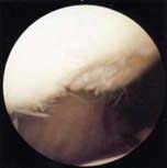
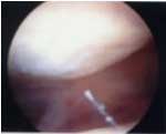
If patellar cartilage deterioration proceeds beyond simple softening and involves fissuring, disintegration and/or outright erosion (see FIGURE 1), the joint surface can become quite irregular and a sensation of grinding or catching may be felt by the patient when extending (straightening) the knee. This is often more pronounced when ascending stairs, or when getting back up from a bent-knee (squat) position. Such symptoms can often be improved by surgery. During an arthroscopic "chondroplasty" procedure the roughened and degenerated articular cartilage tissue behind the patella is shaved down, re-contoured and evened out to the maximum extent possible or practical, using arthroscopic instruments (see FIGURE 2). Laser or radio frequency (r.f.) electro-thermal ablation "sculpting" techniques can also be employed, but extreme care must be taken not to allow excessive heat build-up within nearby healthy cartilage or the underlying bone, as this can cause localized cartilage coagulation, or worse, bone death (avascular necrosis), with potentially catastrophic results. I once encountered a patient treated by another surgeon, in whom a simple, outpatient laser chondroplasty resulted in enough bone death within the patella to ultimately require its complete surgical removal (patellectomy)!
After patellar surgery a patient's knee and leg may temporarily behave as if they are "in shock". The knee joint may remain swollen and irritable for quite some time, and the leg may lose quite a bit of thigh muscle tone and strength. Sometimes special physical therapy regimens that involve electrical muscle stimulation and biofeedback are required to gradually "re-educate" the dysfunctional leg muscles, teaching them how to contract fully again so that they may then be strengthened through routine resistance exercise. Despite such difficulties, if a patient's knee pain is alleviated by the surgery, near-complete restoration of muscle function may be possible following a diligent rehabilitation regimen. Arthroscopic treatment of chondromalacia patella, with or without lateral retinacular release or other more major procedures, can produce gratifying results, but does not represent a total "cure" for the problem. Once articular cartilage has become degenerated, simply shaving away the worst of it does not really restore a joint to normalcy. Sometimes the procedure is palliative, at best, and subsequent surgery and/or specialized brace treatment may be needed. Despite recent advances in surgery, physical therapy, medications and knee bracing, the problem of the stubbornly painful patellofemoral joint is one that orthopedic surgeons have yet to solve, short of prosthetic joint replacement.
|
|||||||||||||||||||||||||