
|
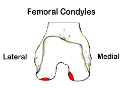
Osteochondritis dissecans is an unusual affliction of human joints that is not rare but also cannot be considered common. The knee is the most frequently affected joint in the body. Curiously, one particular location on the medial femoral condyle (inner aspect of the lower end of the thigh-bone) is where the majority of knee OCD lesions are found (see FIGURE 1).
The next most common site is the posterior aspect (rear portion) of the lateral (outer) femoral condyle (see FIGURE 1), followed by rarer forms in the patella (kneecap) and upper tibia ("shinbone"). OCD begins in childhood and is therefore most commonly seen in teenagers and young adults. Severe (large) OCD lesions that remain unhealed can ultimately wreak havoc on a knee joint, with long-term arthritic consequences that may require joint replacement surgery. Osteochondritis dissecans is a truly mysterious joint disease
process that was studied by 19th century pathologists and given
its name because it was considered an inflammatory (the "itis" in osteochondritis refers to inflammation)
condition of articular (joint surface) cartilage and the underlying
(subchondral) bone. (Note: "osteo" refers to bone and "chondro" refers
to cartilage). The
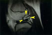
disease causes a section of joint surface cartilage and the bone
beneath it to loosen and separate (by way of a gradual dissection
process, hence the "dissecans" in OCD's name) from the
main or "parent" bone structure such as a femoral condyle (see FIGURE 2).
The actual cause of OCD is unknown. There are three
theories regarding the origin of OCD, the first being that
it is the result of traumatic impact injuries delivered
to the adolescent joint surface, either acutely (suddenly) or
chronically (repetitively) over time. This view considers OCD
to represent a fracture of sorts. The second theory is
that the separating osteochondral (bone and cartilage) fragment
starts out as a small, anomalous (aberrant or extra), independent
zone of ossification (bone formation) during early adolescent
skeletal growth that simply fails to fuse (merge) with the main
ossification center as the bone matures. This ultimately leaves
the bone tissue in that extra ossification center (the "ossific
nucleus" or subchondral ossicle of OCD) isolated and without
a blood supply, depriving it of oxygen and nutrients. The bone
ossicle may then shrink and atrophy, thereby undermining the overlying
joint surface and making the involved segment of articular cartilage
(with attached ossicle) subject to gradual loosening and separation
from the parent bone. The third theory is that normal bone
underlying a region of articular joint cartilage somehow loses
its circulation suddenly and therefore dies, similar to
the way a localized area of heart muscle dies when a blood clot
cuts off its circulation in the case of a myocardial infarct (heart
attack). While my personal experience with many cases of OCD over
the years has led me to believe that the anomalous ossification
center theory is the most likely of the three to be correct,
no one knows for sure. |
|
|
|
|
|
|