Methods of ACL Reconstruction
Completely torn ACLs almost never heal on their
own, and unlike tears in some other knee ligaments, cannot be
stitched back together very effectively. The injured ACL must
almost always be surgically rebuilt, or "reconstructed",
using a replacement ligament (tendon autograft or allograft).
Autograft tissue is harvested from the patient's own body
whereas allograft tissue is obtained from a tissue bank.
Many orthopedic surgeons who perform ACL reconstruction have one
preferred surgical method or technique, which they learned
during their surgical training, and thus feel most comfortable
with. They therefore use this method in the great majority of
their cases, regardless of the patient's age, sex, activity level
or occupation. This is not the approach that we take at The
Knee and Shoulder Centers. We feel comfortable performing
all of the commonly used methods of anterior cruciate ligament
reconstruction and use a variety of different tendon grafts as
an ACL replacement. Each person with a torn ACL represents a unique
situation that calls for surgical decision-making customized to
the particular context of that patient's case.
For example, a time-tested and very commonly performed method
of ACL reconstruction that utilizes an implanted ligament graft
composed of the middle third of the patient's own patellar tendon
(see FIGURES 7a, 7b) typically provides
excellent restoration of knee stability 90 or more percent of
the time, but is sometimes a difficult procedure to recover from.
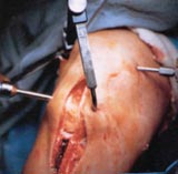 |
FIGURE 7a
- This photo demonstrates the patellar tendon autograft
method of ACL reconstruction, whereby the middle 1/3 of
this frontal knee tendon, with a bone plug at either end,
is first excised and then re-implanted inside the knee where
the ACL originally was. The defects in the patellar tendon,
patella and tibia left from the graft "harvesting"
procedure gradually fill in with scar and repair tissue.
|
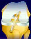 |
FIGURE
7b - Diagrammatic illustration of how a patellar tendon
graft is surgically implanted into the knee where the original
ACL was located. Two screws hold the (bone plug) ends of the
new ACL in place.
|
In our experience, this surgical method poses a higher risk for
unwanted side-effects such as patellar tendinitis, patellar pain,
joint stiffness, internal scarring, and an inability to kneel
on firm surfaces (any or all of which could be permanent).
For this reason, we view this particular procedure as being more
ideally suited to younger (under 25), high-demand athletes who
are not likely to be called upon to kneel on hard surfaces in
an occupational setting. Patellar tendon autograft ACL reconstruction,
done in the wrong patient, has a significant chance of leading
to an unhappy result. We are of the opinion that an individual
over 25 with an acute (recent) anterior cruciate ligament injury
that has not yet resulted in a highly unstable joint, and who
later may be required to do more kneeling activity in an occupational
setting than cutting or pivoting in an athletic environment, is
better suited for other methods of anterior cruciate ligament
reconstruction. These alternate methods utilize either two of
the patient's own accessory hamstring tendons (semitendinosus
and gracilis) as an ACL graft (see FIGURE
8), a part of the patient's own quadriceps tendon from
the front of the lower thigh, or an allograft tendon specimen
obtained from a tissue bank (see FIGURE
9).
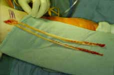 |
FIGURE
8 - This photo demonstrates how two accessory (thus
relatively expendable) hamstring tendons can be retrieved
from the thigh through a 1½ inch long incision, made
just below the knee. These tendons are then doubled or tripled-over,
and fashioned into an ACL graft that is arthroscopically implanted
into the knee at the site where the original ACL was located. |
|
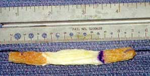
FIGURE
9 - Photo demonstrating an "allograft"
bone/patellar-tendon/bone specimen that has been obtained
from a certified tissue bank. The photo shows the specimen
after it has been cut down to size and fashioned into a
new ACL. The white, middle section becomes the new ligament
and the bone plugs at either end serve as anchors that become
imbedded in the femur and tibia.
|
 |
These alternate techniques leave the patient's
own patellar tendon untouched and rarely produce sensitive
areas in the front of the knee to be bothered later on by floor
contact when kneeling. While there is no surgical knee procedure
that poses zero risk of an unhappy or frankly failed outcome,
these alternate methods are often better tolerated by many patients
as compared with ACL reconstruction using their own patellar tendon.
There are also various specific technical advantages and disadvantages
to each particular surgical method and/or ACL graft when considered
in the context of a patient's exact clinical circumstances. Taking
into account these various details, educating the patient about
them and then jointly arriving at a decision as to how to proceed,
is our preferred approach to the problem of the ruptured anterior
cruciate ligament. Our goal is to subject a patient's knee
to no more surgical stress than is necessary to
achieve the desired result.
|